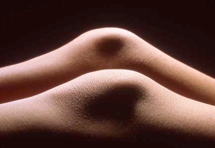
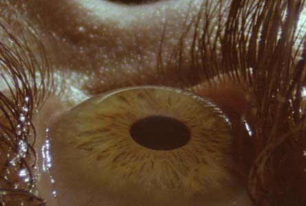
My macro and micro photography of the body involves viewing it from an unusual and hopefully interesting perspective. Some of this can be seen in the Archive Showreel. However, there are many things, especially inside the body, that really cannot be filmed. Then we enter the world of special effects; simulating the environment using real tissue rather than models or CGI.
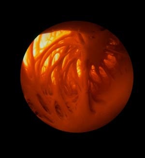
The picture to the right is taken using an endoscope inside the atrium of a heart. The view shows the muscle bands in the wall of the auricle. The heart appears to beat in movies because it is pressurised with saline whilst being given external cardiac massage.
Simulating reality using real biological subjects is not always possible. Inevitably this leads to building models of some aspects of the human body and human development. This also allows us to work at a bigger scale and avoid some photographic problems such as limited depth of field. It creates a experience where the viewer feels that they are really inside the body observing life as it happens.
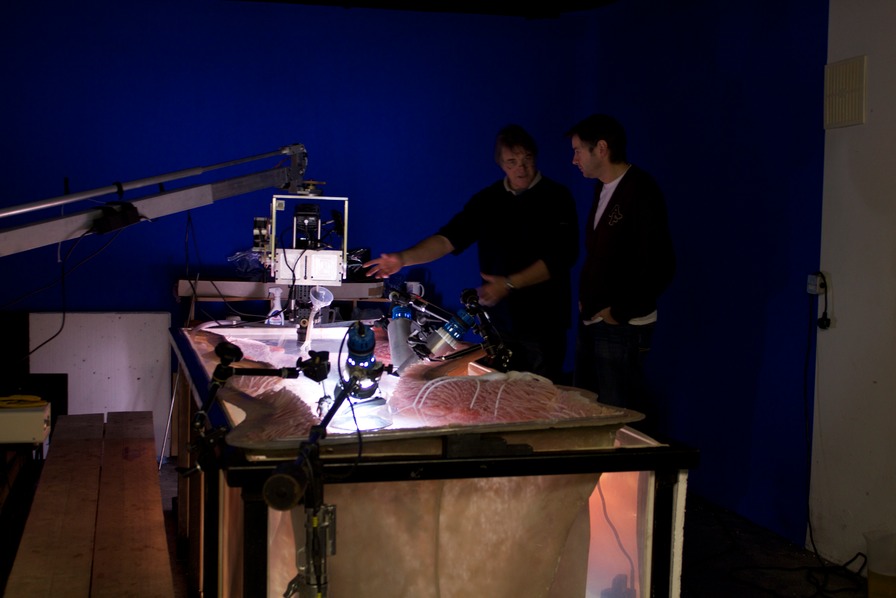
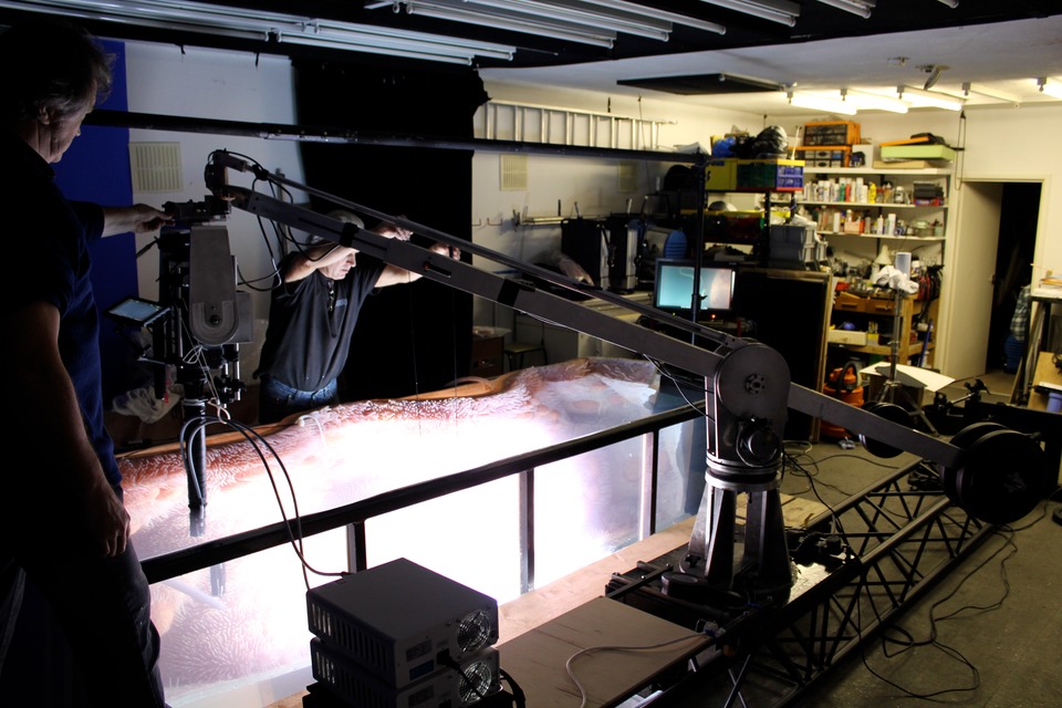
The Discovery film ”Birth” is an example. It involved very large models including a three metre fallopian tube set complete with sperm cells and cilia. The picture here shows the set full of water whilst I am discussing a shot with director Alex Hearle.
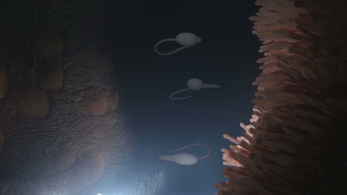
More detail is seen in the next picture that shows two model sperm cells “swimming" along the tank. The sperms were operated by puppeteers. The poles had to be painted out later.
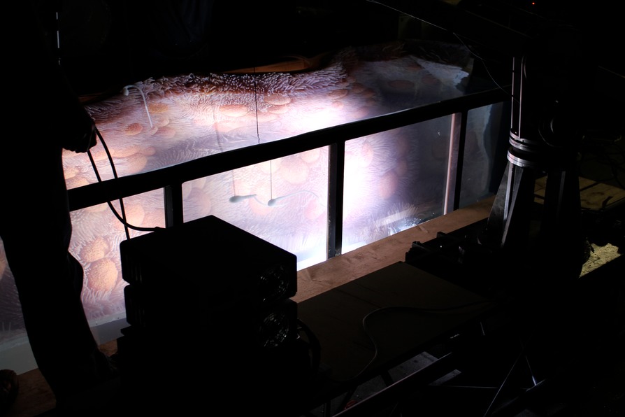
The third picture in the sequence shows the rig that carried the camera connected to a borescope in a waterproof sheath. The whole rig ran on a track so that it could move up and down the tank whilst filming. The boxes next to me in the foreground are metal halide light sources for the fibre optic cables attached to the camera borescope. These were not the main lights but were there to provide a fill to the main lights shining through the walls of the tank and the water surface.
The final shot is seen in the picture below. Note that there is only one wall in the model fallopian tube and yet 2 in the image. The opposite side of the fallopian tube was, in fact, a reflection in the wall of the perspex tank. Sometimes the simple things work best!
Numerous other large scale models have been filmed including: human and animal foetuses and embryos, ears, brains, arteries and skin. Stills of some of the human and animal baby models in their womb sets can be seen in the Gallery.
A Showreel of some of the microscopy and macro photography as well as some of the most recent movie work on the National Geographic feature: "Cradle to Grave” can be found in the Showreels section.